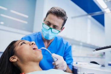
Sialography
Sialography is a radiographic imaging technique used to visualize the salivary glands and their ductal systems. This procedure employs contrast media, which are substances that enhance the visibility of internal structures on X-ray images. By injecting a contrast agent into the salivary ducts, sialography provides detailed images that assist in diagnosing various conditions related to the salivary glands.
Anatomy of the Salivary Glands
The human body has three major pairs of salivary glands: the parotid, submandibular, and sublingual glands. In addition, there are numerous minor salivary glands distributed throughout the oral cavity.
Parotid Glands: Located near the jaw and primarily responsible for producing serous (watery) saliva, which aids in digestion.
Submandibular Glands: Found beneath the jaw, these glands produce both serous and mucous saliva.
Sublingual Glands: Situated under the tongue, they mainly produce mucous saliva, which lubricates food and aids in swallowing.
Saliva plays a crucial role in oral health, helping to break down food, neutralize acids, and maintain a balanced oral environment.
Indications for Sialography
Sialography is typically indicated for various conditions affecting the salivary glands, such as:
Sialolithiasis: The formation of stones within the salivary ducts, leading to obstruction and inflammation.
Sialadenitis: Inflammation of the salivary glands, often due to infection or blockage.
Tumors: Both benign and malignant growths can occur in salivary glands, and imaging is essential for assessment.
Congenital Abnormalities: Some individuals may have structural anomalies in their salivary glands that can be evaluated through sialography.
The Procedure
The sialography procedure typically involves several steps:
- Preparation: Prior to the procedure, patients may be advised not to eat or drink for a few hours. This is to ensure that the salivary glands are active, which enhances the effectiveness of the imaging.
- Injection of Contrast Material: A healthcare professional will insert a small catheter into the duct of the salivary gland being examined. A contrast agent, often a water-soluble iodine-based solution, is then injected through this catheter. This agent will outline the salivary ducts, making them visible on X-ray images.
- Imaging: Once the contrast material is injected, a series of X-ray images are taken. These images will show the outline of the salivary ducts and any abnormalities present, such as stones, blockages, or tumors.
- Post-Procedure Care: After the imaging is completed, the catheter is removed, and patients are generally allowed to resume normal activities. Some may experience mild discomfort or swelling, but serious complications are rare.
Risks and Considerations
While sialography is generally considered safe, there are some potential risks and side effects to be aware of:
Allergic Reactions: Some individuals may have an allergic reaction to the contrast material. It is important to inform the healthcare provider of any known allergies prior to the procedure.
Infection: Although rare, there is a small risk of infection at the injection site or within the salivary glands.
Discomfort: Patients may experience temporary discomfort during the injection of the contrast material or when the glands are stimulated to produce saliva.
Interpretation of Results
The images obtained from sialography are interpreted by a radiologist or a specialized dentist. The findings can provide valuable information regarding the presence of stones, blockages, or tumors, enabling appropriate treatment plans to be developed.
Conclusion
Sialography is a vital diagnostic tool in the field of dentistry and oral medicine. By providing clear images of the salivary glands and their ducts, it aids in the diagnosis and management of various conditions affecting salivary function. Understanding this procedure can help individuals appreciate the importance of maintaining salivary health and seeking timely treatment for related issues.
Discover affordable dental treatments with Dr. BestPrice!
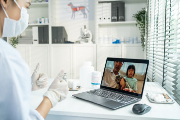
Teledentistry revolutionizes oral healthcare by connecting patients with dental professionals virtually, improving access while reducing costs—discover how remote dental consultations are reshaping the industry.
Discover how this overlooked bodily fluid acts as your mouth's personal bodyguard, fighting bacteria, rebuilding teeth, and maintaining oral health around the clock.
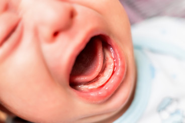
From teething troubles to first dental visits, uncover the truth behind common baby teeth myths and learn how to protect your child's precious smile.