Periapical Radiograph
A periapical radiograph, often referred to as a periapical X-ray, is a specific type of dental X-ray that captures the entire tooth—from the crown, which is the visible part of the tooth above the gum line, to the root, which is embedded in the jawbone. This imaging technique is crucial for diagnosing a variety of dental conditions, particularly those affecting the tooth roots and surrounding bone.
Purpose
The primary purpose of a periapical radiograph is to provide detailed images that assist dental professionals in diagnosing issues related to the teeth and surrounding structures. These X-rays are particularly useful for identifying:
Dental Caries (Cavities): Periapical radiographs can reveal the extent of decay that might not be visible during a clinical examination.
Periapical Pathologies: These include infections at the root of the tooth, often indicated by dark areas around the root in the radiograph.
Bone Loss: The images can show changes in the bone surrounding the tooth, which may indicate periodontal disease or other conditions.
Tooth Development Issues: In children, these X-rays can help monitor the growth and development of teeth.
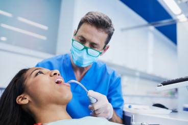
Periapical radiographs are taken using a small X-ray film or digital sensor that is positioned in the mouth. The film is placed close to the tooth being examined, and an X-ray machine directs a controlled amount of radiation toward the sensor. The image produced shows the structures of the tooth and surrounding bone in detail.
Types of Periapical Radiographs
Standard Periapical Radiographs: These typically capture one or two teeth in detail, allowing for focused examination.
Multiple Periapical Radiographs: In some cases, several periapical films may be taken to provide a comprehensive view of multiple teeth in a specific area.
Interpretation of Images
The interpretation of periapical radiographs requires specialized training. Dental professionals examine the X-ray for various indicators of health and disease. Key aspects looked for include:
Root Canal Anatomy: The shape and integrity of the root canal system are assessed.
Bone Density: A healthy bone should appear dense and without dark spots. Dark areas may indicate infection or bone loss.
Cysts or Tumors: Periapical radiographs can reveal the presence of cysts, tumors, or other abnormalities in the jawbone.
Safety and Risks
While periapical radiographs involve exposure to radiation, the levels used are minimal and considered safe. Modern dental practices employ digital radiography, which reduces radiation exposure even further. Dental professionals take precautions to limit exposure, especially in vulnerable populations such as children and pregnant individuals.
Advantages
High Detail: Periapical radiographs provide a clear view of the tooth's structure, which is essential for accurate diagnosis and treatment planning.
Cost-Effective: Compared to other imaging techniques (like CT scans), periapical X-rays are relatively inexpensive and accessible.
Quick Procedure: The process of taking a periapical radiograph is typically quick, often taking just a few minutes.
Limitations
Despite their advantages, periapical radiographs have limitations. They provide a two-dimensional view of a three-dimensional object, which may lead to misinterpretations. Additionally, they may not capture all issues if the area of interest is not included in the image. Therefore, dentists may use other imaging modalities (like panoramic radiographs or cone-beam computed tomography) in conjunction with periapical X-rays for a more complete assessment.
Conclusion
In summary, a periapical radiograph is an essential diagnostic tool in dentistry, providing critical insights into the health of teeth and their supporting structures. By revealing detailed images of the tooth roots and surrounding bone, periapical radiographs help dental professionals diagnose conditions that might otherwise go unnoticed. Understanding this imaging technique can empower individuals to engage in informed discussions with their dental care providers regarding treatment options and oral health strategies.
Explore

Teledentistry revolutionizes oral healthcare by connecting patients with dental professionals virtually, improving access while reducing costs—discover how remote dental consultations are reshaping the industry.
Discover how this overlooked bodily fluid acts as your mouth's personal bodyguard, fighting bacteria, rebuilding teeth, and maintaining oral health around the clock.
From teething troubles to first dental visits, uncover the truth behind common baby teeth myths and learn how to protect your child's precious smile.